Top Apps
Remote Control for TV - All TV
الدولار اليوم في مصر بكام؟
Files by Google
شاهد Shahid
DJ Liker
Videocash
TurboSpeed: Game Mode FPS
OfferGoose-Land Your Dream Job
Notta-Transcribe Audio to Text
PDF Reader: Edit & Convert PDF
دعاء العمرة والطواف والسعي
الأدعية اليومية للأطفال
Psiphon: Fast and Secure VPN
Binary Code Translator
2Accounts - Dual Apps Space
Apps Home
All Rights Reserved © Apps Home 2025

VOKA 3D Anatomy & Pathology
Discover the World of Pathology in 3D
Factory of innovations and solutions LLC
Review (4.1)
Reviews
+2K
Downloads
+100K
Security
Safe


3D Pathology Experience: A Revolution in Medical Learning and Application
In the ever-evolving world of medical science, the integration of modern technology into learning and practice has brought about paradigm shifts in how we perceive and interact with human anatomy and pathologies. The advent of 3D technology in pathology is one such remarkable breakthrough. This immersive learning experience not only enhances understanding but also reshapes educational methodologies and clinical applications.
The Essence of 3D Pathology
The concept of 3D pathology revolves around creating medically accurate, three-dimensional models of human anatomy and associated pathologies, including some that are rare. These models provide a comprehensive, detailed view, unparalleled by traditional two-dimensional textbooks and diagrams. They are instrumental in depicting complex structures and relationships within the human body, offering learners and medical professionals an encyclopedic view of various medical conditions.
Applications in Medical Education
For medical students, the 3D Pathology Experience offers an invaluable resource for understanding complex anatomical structures and relationships. The ability to view organs and systems from any angle, zoom in on specific pathology, and study the interplay between healthy and diseased states provides a deeper insight into human biology. This tool is a game-changer for those studying human anatomy, whether for exams or practical applications, especially when it comes to visualizing intricate details such as vascular pathways, musculoskeletal nuances, or organ-specific conditions.
Benefits for Lecturers and Researchers
Lecturers and researchers benefit from 3D pathology by employing these models in both online and offline lectures and presentations. The interactive nature allows educators to demonstrate real-life clinical scenarios, enhancing the learning experience by providing a visual context that is more engaging and easier to comprehend than static images. Research professionals can utilize these models to visualize hypothetical pathological scenarios, aiding in the exploration of new medical treatments and understanding pathogenesis.
Clinical Implications and Patient Communication
For clinicians, the ability to utilize three-dimensional models in patient consultations can greatly enhance communication and understanding. By providing a clear visualization of a patient's condition, doctors can explain surgical procedures, treatment strategies, and rehabilitation processes more effectively. This not only improves patient comprehension but also empowers them to make informed decisions about their healthcare.
VOKA Anatomy Pro: A Vanguard in 3D Pathology Visualization
Among the innovations leading this field is a comprehensive atlas that offers a rich repository of 3D models. Designed to be used anytime and anywhere, this mobile atlas provides a versatile tool for medical professionals and students. Each model is meticulously developed in collaboration with medical experts, ensuring accuracy and reliability. The models are based on actual DICOM data from CT and MRI scans and are verified by a medical advisory board, ensuring they meet clinical standards.
Features and Accessibility
The applications are designed to be lightweight and accessible, making them suitable for a wide range of devices, whether on mobile phones or desktops. Key features include the ability to zoom, rotate, isolate anatomical structures, and view models from any angle. These functionalities enable users to tailor their learning or clinical presentations to specific needs, enhancing both understanding and application of medical knowledge.
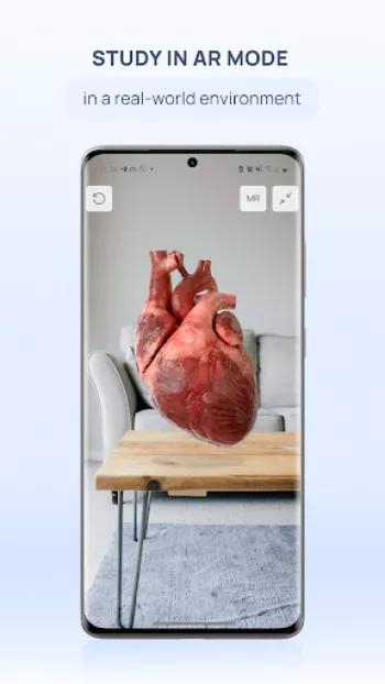
Augmented Reality and Future Directions
Augmenting the experience, the AR mode lets users overlay virtual 3D models within real-world settings, fostering an immersive learning environment. This capability is particularly beneficial for detailed studies of complex structures such as the circulatory system, cranial nerves, and more. As technology evolves, the integration of even more advanced features like 4D and 5D modeling promises to further enhance the capabilities and applications of 3D pathology in medical sciences.
Conclusion
The 3D Pathology Experience represents a significant advancement in medical technology, providing numerous advantages to educators, students, clinicians, and patients alike. By offering an immersive, interactive, and accurate representation of human anatomy and pathologies, it bridges the gap between theoretical learning and practical application, ensuring a more comprehensive understanding of medical complexities. This tool is not only revolutionizing medical education and research but also enhancing clinical practices worldwide.
To explore and experience these models, you can find them on various platforms for diverse operating systems.
شاركنا رأيك
بريدك الالكتروني لن يتم نشره.


All Rights Reserved © Apps Home 2025
Muniza Ansari
Amazing, amazing app. Extremely detailed and accurate. One of the finest apps I've come across so far. It's fueling my love for anatomy, explaining...
Mahesh Sankhala
One of the best 3d anotomy app. Great for education for children, adult and health care professionals. And ability to show /hide individual section...
Krishna
very good app for anatomy and pathology. but great things are they r not taking any subscription for full app uses. it's totally free. thanks for it.
Tyson
bestest free anatomy application in the entire world. Deserve more. Free anatomy of osteology is often provided free of cost in other applications ...
Giorgi Shvelidze
This app helped me learn more about anatomy, structures and organ's functions. It's completely free (from what I've seen and discovered) and it's a...