Top Apps
Goodreads: Book Reviews
Fajr: Fajr Alarm, Prayer Times
BlackHole
Grand Mobile:RP Life Simulator
Question.AI - Chatbot&Math AI
AG Auto Clicker-Auto Tap
ElevenReader - Text to Speech
Plex: Stream Movies & TV
Google Tasks
CapCut - Video Editor
iLovePDF: PDF Editor & Scanner
Coinbase: Buy BTC, ETH, SOL
FlashGet Kids:parental control
STARZ ON
ExpertOption - Mobile Trading
Apps Home
All Rights Reserved © Apps Home 2025

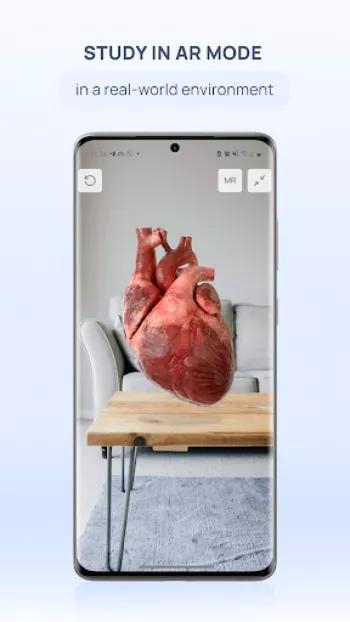
VOKA 3D Anatomy & Pathology
Discover the World of Pathology in 3D
Factory of innovations and solutions LLC
Review (4.1)
Reviews
+2K
Downloads
+100K
Security
Safe


Exploring the Revolutionary Approach of 3D Pathology
The modern field of pathology is undergoing a significant transformation with the introduction of 3D visualization techniques that are redefining diagnostic capabilities and educational methods. The concept of 3D pathology involves creating lifelike, three-dimensional models of human anatomy and its pathologies, which can be manipulated and observed from various angles in ways that traditional two-dimensional imaging simply cannot achieve. This technological advancement is empowering clinicians, educators, and students alike by providing a comprehensive view of the human body's intricate structures. With the help of programs like VOKA Anatomy Pro, these models offer unparalleled accuracy and detail because they are based on DICOM data from CT and MRI scans, ensuring that they reflect true-to-life anatomy. This is crucial because accurate, detailed visualization allows for better diagnosis and a deeper understanding of complex medical conditions. By using 3D models to study pathologies such as congenital heart defects or acquired heart diseases, medical professionals can gain more insight into how these conditions develop and progress. Furthermore, by simulating the pathologies in a virtual environment, researchers can test and evaluate new treatment methods in a risk-free setup. This revolution in how anatomy is visualized and understood is not merely about looking at pretty pictures; it reflects a profound shift in training methodologies and patient care standards. Through realistic 3D anatomy modeling, medical students are now able to study and visualize the entire human body in staggering detail, drastically improving their memorization and comprehension of complex anatomical structures. As a result, students become more adept at recognizing pathologies and understanding their implications within different biological systems. This revolutionary approach does not discriminate; it covers a broad range of anatomical particulars, including internal organs, circulatory systems, and even nuanced cases in otorhinolaryngology and gynecology, offering a wide-reaching impact on medical education and practice.
The Technical Foundation of 3D Pathological Models
Constructing 3D pathological models involves an intersection of advanced imaging techniques, software engineering, and medical expertise. VOKA Anatomy Pro exemplifies this by integrating actual CT or MRI scan data into fully interactive models. The process starts with capturing high-resolution images through advanced medical imaging technologies that serve as the raw data. These scans are rich in detail and provide a cross-sectional view of the body's internal composition. From there, software engineers and medical experts collaborate to 'stack' these 2D images, converting them into coherent 3D structures. This software-intensive process applies numerous algorithms to ensure that each voxel (a pixel with volume) in the data reflects the correct tissue type and morphological structure. What renders this system revolutionary is its accuracy and flexibility; users have the ability to dissect, segment, and annotate various anatomical features, exploring them from any angle or depth. This manipulability extends the application of 3D models beyond mere visualization—they become tools for diagnostic analysis, medical training, and surgery preparation. For example, a surgeon planning a complex procedure can use a 3D model for pre-operative evaluation, studying every nuance to plan their approach. Even pathologies can be isolated and examined without any interference from surrounding tissues, a feature that makes next-generation 3D models indispensable in a clinical setting. Beyond diagnostics and treatment, these models serve as an unparalleled educational aid that allows students to interact and engage with lifelike anatomical structures. 3D printed models derived from these digital constructs further bridge the gap between theoretical knowledge and practical application, offering tangible references for hands-on learning. Thus, the technical prowess underpinning 3D pathological models lies at the core of not only advancing medical science but also facilitating a deeper comprehension of human health dynamics.
Education and Training in the Era of 3D Modeling
Incorporating 3D models into medical education is transforming the landscape of learning and professional training. Historically, medical education relied heavily on textbooks and fixed slides, methods that, while informative, limited a student’s ability to interact with the material. However, with the advent of 3D pathology, learners can now experience dynamic and interactive lessons that significantly enhance understanding and retention. Applications like VOKA Anatomy Pro offer an agile learning platform where users can zoom in on areas of interest, rotate models in 360 degrees, and isolate or hide structures to study their relationships in isolation. This approach encourages an interactive learning environment where students can actively engage with the subject matter. For educators, this technology promises to transform lesson delivery by facilitating more engaging lectures and practical sessions. Academics can use these models to demonstrate complex anatomical interactions and contextualize theoretical knowledge with visual evidence. Virtual labs can emulate real-world scenarios, providing students with experiential learning opportunities that traditional methods cannot offer. The opportunity to explore the pathology in three dimensions helps students visualize how body systems and structures are affected, promoting a deeper level of analytical thinking. Moreover, 3D pathology models support various teaching methods—such as problem-based learning and team-based scenarios—enabling a diversified educational approach. They provide the scaffolding upon which students can build their expertise, from basic anatomical structures to more complex pathological phenomena. This educational paradigm shift ultimately equips medical students with the skills needed to meet the demands of an ever-evolving healthcare landscape, where robust anatomical understanding plays a critical role in delivering patient-centered care. By transcending the limitations of traditional education, 3D pathology integration represents a leap forward in the evolution of medical training, providing a versatile, effective means of knowledge dissemination.
Real-World Applications and Implications of 3D Pathology
The transition from theoretical applications to real-world integrations of 3D pathology marks a transformative phase with wide-reaching implications for healthcare. In clinical settings, 3D models are being leveraged to enhance diagnostic accuracy, facilitate surgical planning, and improve patient education. Surgeons often face challenges in visualizing complex anatomical structures, particularly in cases involving rare or multifaceted pathologies. With 3D modeling, these challenges can be mitigated by providing a comprehensive, manipulable view of the surgical site. For example, a neurosurgeon can review a patient's brain model pre-surgery to determine the best path to a tumor, minimizing risk and improving outcomes. These models also support minimally invasive surgical techniques by offering better insights into the spatial relationships of anatomy. For patients, 3D models serve an educational purpose as well. Visual aids help in the understanding of diagnoses, proposed treatments, and surgical risks, thus enhancing communication between healthcare providers and patients. With the ability to see a model of their own anatomy, patients can make more informed decisions about their care. Additionally, the integration of augmented reality with these models, as seen in VOKA Anatomy Pro, adds a layer of real-world interactivity by superimposing anatomical models onto the user’s environment, offering further insight into human anatomy in motion. Beyond curative applications, these models are beneficial in tracking disease progression over time, aiding in personalized medicine approaches wherein treatments are tailored based on an individual's unique anatomical structure. 3D pathology’s potential to streamline medical procedures and improve healthcare delivery exemplifies its indispensable contribution to modern medicine. It indicates a future trend toward more precise and patient-centered care, where the benefits of advanced visualization are fully realized.
Launching Your 3D Pathology Journey: Tools and Accessibility
Breaking into the realm of 3D pathology exploration is now more accessible than ever, thanks to user-friendly platforms like VOKA Anatomy Pro, which are designed for widespread use across various devices. As these tools become integral to both medical education and professional practice, they underscore the necessity of providing a seamless, cross-platform experience. Key to this accessibility is the availability of the application on diverse operating systems, from mobile to desktop platforms. Users can effortlessly download and utilize VOKA Anatomy Pro across devices, enabling constant access to an expansive library of 3D anatomical models and medical resources, even in environments with limited internet connectivity. For Android users, for example, the journey begins with a simple download and installation process, bringing the world of 3D models to one’s fingertips. These applications are lightweight yet powerful, engineered to function efficiently without demanding significant hardware resources. Whether using a smartphone for on-the-go learning or a desktop setup for more in-depth exploration, the adaptability of VOKA Anatomy Pro ensures its utility in any context. Furthermore, the application supports several languages, making it an inclusive tool for a global audience. For users interested in exploring these innovative tools, starting is as simple as selecting your device's dedicated option: Download for Android. The journey into 3D pathology exploration promises not just practical learning but a transformative understanding of human biology that is as engaging as it is enlightening, paving the way for a future where technology and medicine are inextricably linked.
Share Your Opinion
Your Email Will Not Be Published.


All Rights Reserved © Apps Home 2025
Muniza Ansari
Amazing, amazing app. Extremely detailed and accurate. One of the finest apps I've come across so far. It's fueling my love for anatomy, explaining...
Mahesh Sankhala
One of the best 3d anotomy app. Great for education for children, adult and health care professionals. And ability to show /hide individual section...
Krishna
very good app for anatomy and pathology. but great things are they r not taking any subscription for full app uses. it's totally free. thanks for it.
Tyson
bestest free anatomy application in the entire world. Deserve more. Free anatomy of osteology is often provided free of cost in other applications ...
Giorgi Shvelidze
This app helped me learn more about anatomy, structures and organ's functions. It's completely free (from what I've seen and discovered) and it's a...